

생체내 공초점 이미징 시스템
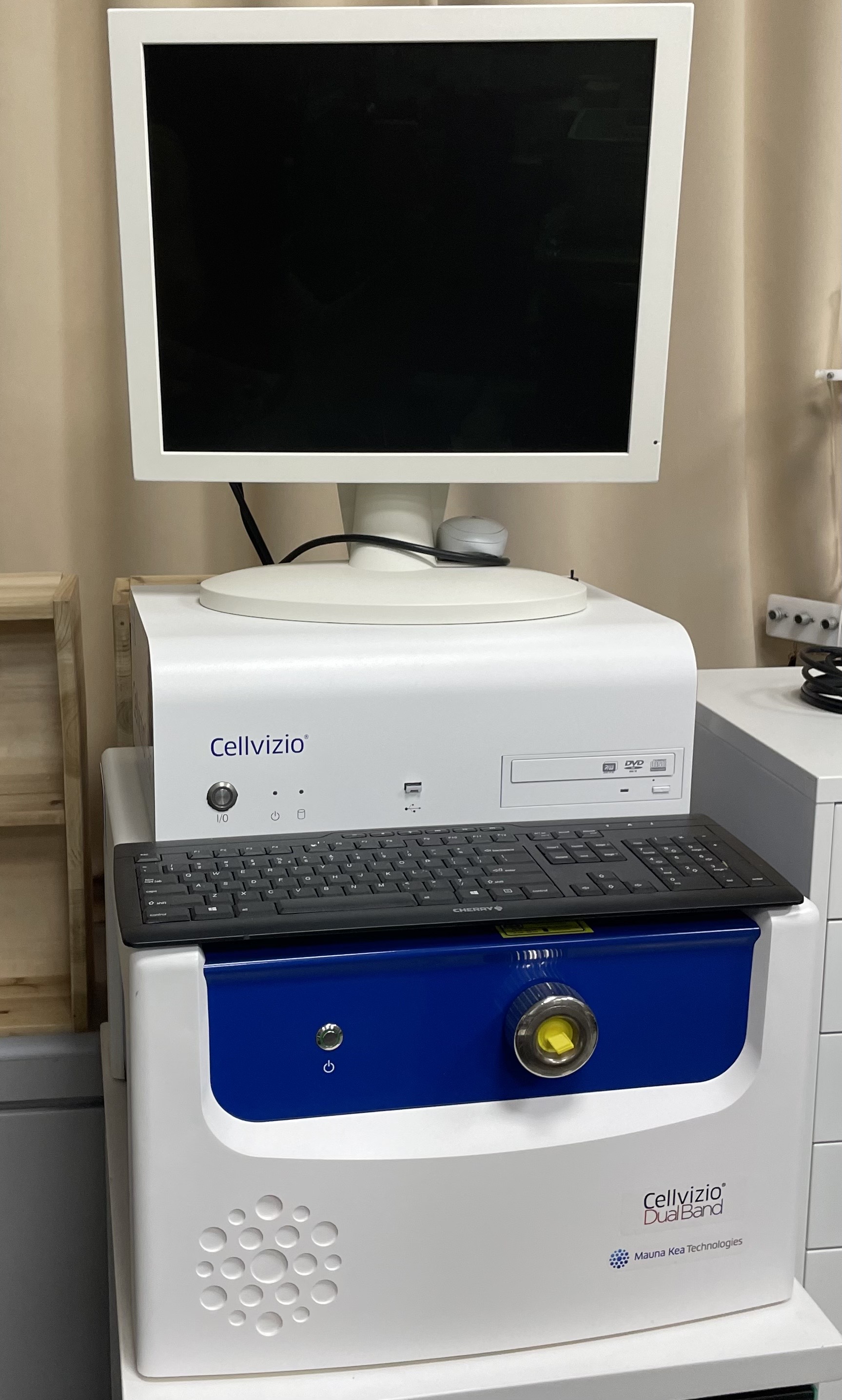
In vivo confocal Imaging systemCellvizio Dual band
기본정보
 |
|
| 등록번호 | NFEC-2016-11-212639 |
|---|---|
| 모델명 | Cellvizio Dual band |
| 제작사 | Mauna Kea Technologies |
| 설치장소 | 5 전기생리실 |
| 구축일자 | 2016-10-17 |
| 전자메뉴얼 | |
| 담당자 | 최준호 |
동영상&장비사진

구성&기능
[구성]
1) Dual Laser Scanning Unit 488 and 660nm dual band excitation/detection
2) Confocal processor & High resolution color monitor
3) Accessories - Foot pedal control
- Probe holder
- Cletop-S box with 1 refill(for probe cleaning)
- Additional caps for ProFlex Microprobes
4) Image acquisition and processing software
5) IC Viewer software 6) Vessel detection software 7) Advanced Mosaicing software 8) ProFlex S1500 probe 9) NeuroPak 10) 3-pack Quantikit (Cleaning and calibration kit) for 488nm
11) 3-pack Quantikit (Cleaning and calibration kit) for 660nm
[성능]
1) Dual Laser Scanning Unit 1. Excitation wavelength 488 & 660 nm 2. Frame rate 9 ? 50 fps 3. Resolution 1.4 um 4. Laser Class 2M
2) Confocal processor & High resolution color monitor 1. Processor: Twol 2.8 GHz Quad-Core Intel Exon 2. External ports: 5 USB, 2 Firewire 400, 2 Firewire 800 3. Graphic board: NVIDA geForece 8800 GT 512MB GDDR3 4. Hard Drive: 320GB(or above), 7200 rpm Serial ATA 3Gb/s 5. DVD: One 16 x SuperDrive 6. Operating system: Leopard 10.5x 7. RAM: 2GB (2X 1GB ECC FB-DDR2 800MHz) 8. High resolution 19“ flat panel color monitor (1280x1024 pixels) 9. Keyboard, mouse and documentation
3) Accessories 1. one foot pedal control for image sequences acquisition and save 2. one ProFlex holder 3. one set of standard operating maunals 4. fuses and cables 5. additional caps for ProFlex microprobes and Cellvizio connector
4) Image acquisition and processing software 1. Image-cell operating software License (Pre-installed on the computer) 2. Calibraton module for Image quality control 3. Acquisition module for real-time acquisition and processing
본 장비는 생체 세포 및 조직 또는 장기에서 목적 단백질의 기능 규명을 위해 형광 염색 처리된 물질을 초 고감도 해상력으로 실시간 분석가능하다. 매우 정교하고 signal이 강한 Laser source를 이용하여 형광 signal을 극대화 하였으며 다양한 confocal micro probe를 통해 surface에서부터 deep tissue까지 관찰 가능하며 최대 1.4 μm의 resolution으로 매우 뛰어난 영상을 관찰 할 수 있다.
